Lipomas

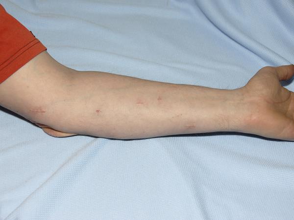
Lipomas are the most common cause for a lump under the skin. They may be single although some people make multiple lipomas.

hurren-lipomas-hero
Lumps

20+ Years

Experience

Lipomas are benign tumours of fat. They are very common and are found all over the body. They are not usually harmful and are not considered pre-cancerous (ie do not turn into cancers). They may go on growing and may be unsightly or uncomfortable. They are usually removed by making a cut over the lipoma and removing it. The wound is then usually closed with dissolving stitches. Some lipomas ‘pop out’ very easily others can be difficult to remove. Removing a lipoma leaves a scar on the skin. Lipomas can grow back (recur).

Some lipomas can be removed using liposuction. The main advantage is that this results in smaller scar and the scar can be placed away from the lipoma.

Summary – Lipomas

Procedure

The operation is usually carried out under local anaesthetic (injection to make the skin numb). Larger lipomas or those lying deeper, such as in muscle, may require a general anaesthetic (asleep). It is usually classified as an outpatient or daycase procedure. You are usually only in the hospital for a short period before and after the procedure. With larger lipomas a surgical drain and an overnight stay in hospital may be necessary.

Excision

Usually a cut is made over the lump which is then removed. If the lump is in the skin then it is usually excised by removing a diamond shaped piece of skin with the cyst in the centre. The diamond shape is then closed with stitches to leave a straight line. Stitches may be absorbable (dissolving) or may need to be removed. On the face usually stitches that need to be removed are used.

Testing (histology)

It is usual practice for all bits of skin or lumps that are removed to be sent to the histology laboratory for analysis to check they are benign. In some instances the diagnosis may be so clear that this is not necessary.

Frequently Asked Questions

A lipoma is a benign tumour of fat. It is made up of fat cells that are filled with fat. Fat cells or lipocytes are part of the body’s energy storage system. A minor malfunction can cause a fat cell to enlarge and reproduce to form a lipoma. Lipomas are not cancers. A cancerous or malignant tumour of fat is called a sarcoma or specifically a liposarcoma.

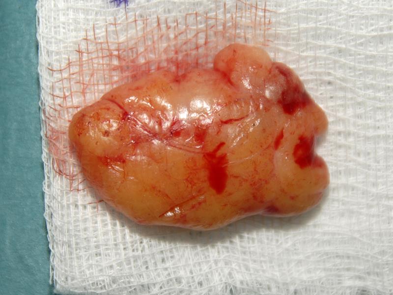
To the naked eye they look like normal fat. In fact even under a microscope they are usually indistinguishable from normal fat. They may be a smooth yellow lump or be made up of lobules.

Scans can be helpful to give more information about the nature of a lump. If the nature of a lump is uncertain then a scan can be helpful. Scans can also be helpful if the precise location of the lump is not clear. Most lipomas are found in the fat under the skin and are usually called ‘subcutaneous lipomas’. Lipomas can develop in the muscles or deeper in the body. Sometimes it is difficult to be sure if what appears to be a small lipoma does not extend more deeply into the body. That is to say it may be the tip of the iceberg. Usually ultra sound scans or MRI scans are the most helpful.

This depends very much on its size and how stuck it may be to surrounding tissues. Many small lipomas sitting just under the skin come out very easily. Others can have lots of lobules that are attached to other structures.

Small lipomas lying just under the skin can be removed with very little pain after the surgery. Larger lipomas especially if positioned more deeply in the body can cause pain after surgery. There is considerable variation between different people and different sizes and sites of lipomas.

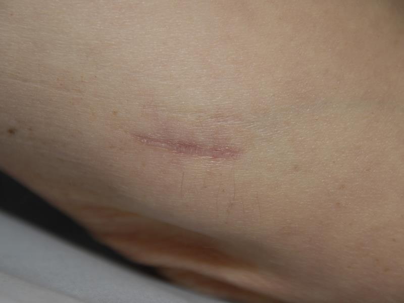
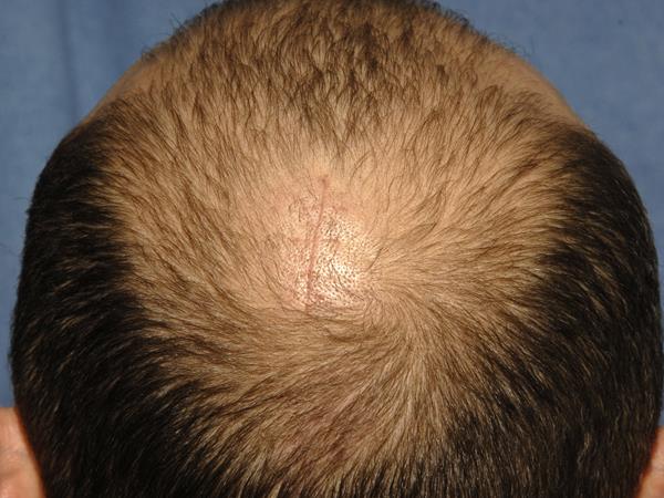
There will usually be a scar over the site of where the lipoma used to be. Depending on how difficult the lipoma is to remove this may be quite short in relation to the lipoma but with more difficult lipomas the scar may be longer than the length of the lipoma.  As the lipoma pushes the normal fat away once the lipoma has been removed it may leave a depression in the skin.

All surgery carries risks. The main risks are:

Bleeding and haematoma

Wounds can bleed after surgery.  A build-up of blood under the skin is called a haematoma. A haematoma may require more surgery to remove it. A haematoma may affect the appearance of the end result.

Infection

A surgical wound can become infected. With surgery on uninfected clean skin this unlikely with less than 1 out of every 25 wounds becoming infected. These will usually settle with antibiotics but it is possible for the infection to form an abscess and then discharge through the wound or require surgery to drain it. Infection progressing to sepsis which is potentially life threatening is very rare in healthy people having skin surgery.

Wound breakdown and delayed healing

Wounds can open up or breakdown. This is most likely to happen if the wound is stressed in the early stages of healing or if it becomes infected. The wound will then usually heal ‘from the bottom up’ and this may take several weeks. The result is usually a wide scar that may be depressed. Some people may want further surgery to try and improve the appearance.

Poor scarring

Most people find the scar from surgery acceptable and worth the benefits of the surgery. The appearance of scar varies a lot from one person to another. Some people may very good nearly invisible scars when others with a similar wound may make an unsightly poor scar. Poorly planned surgery and complications like infection may well lead to poor scars. For other people a genetic tendency to make hypertrophic or keloid scars may result in a poor appearance of a scar. The section on the website about scarring has more details on poor scars.

Deformity after surgery

When a large lipoma is removed from under the skin it may result in a depression in the skin. The lipoma having pushed away the normal fat and tissues under the skin. Once the lipoma is removed there is an inevitable depression in the skin. These usually improve with time but to a degree are permanent.

Recurrence of a lipoma

If a lipoma is completely removed it is unlikely to recur. If part of the lipoma is left behind when it is removed surgically then recurrence is much more likely.

If you have made one lipoma you are more likely to make more lipomas in the future.

Risks of not having a lipoma assessed or treated

There are risks of treatment but there are also risks of not having treatment. An untreated lipoma make enlarge and become more difficult to treat. A ‘lipoma’ that has not be assessed may turn out to be more serious than a simple lipoma.
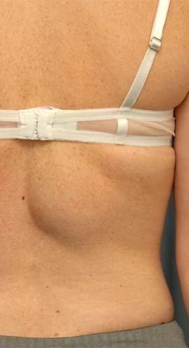
Lipomas are benign tumours. They are an abnormal growth of the body’s fat cells. They are benign which is to say that the cells are not so abnormal as to be considered malignant which means cancerous. Cancerous cells invade aggressively into the tissues and structures around them and can spread to other parts of the body at which point they are called a metastasis. Cancerous cells go on multiplying out of control. Lipomas can grow into structures next to them but usually do so by following tissue plains rather than invading. Lipomas do not spread to other parts of the body in the blood stream. That is to say they do not metastasise.  Lipomas may usually grow slowly and often stop growing. Sometimes they can reach quite large sizes but their growth is not normally considered out of control in the way of a malignant or cancerous* tumour.

Lipomas are not considered pre-malignant. That is to say that they are not considered at significant risk of becoming cancerous. The risk of small lipomas lying under the skin of becoming malignant is so low as to not merit removing them.

It is possible to have malignant or cancerous tumours of fat. These are called liposarcomas not lipomas. They vary from relatively nonaggressive tumours to highly aggressive malignancies that metastasise around the body. These are very rare compared with lipomas. Liposarcomas generally grow more rapidly, are larger and are more likely to be tender or painful.

There is a tumour called an ‘atypical lipoma’ or ‘atypical lipomatous tumours’. These are borderline malignant and have some more malignant features than lipomas. In the past they were called well differentiated liposcarcomas. However, calling them a type of liposcarcoma was considered misleading as they do not behave in a typically malignant way and they are now called atypical lipomatous tumours.

Atypical lipomas have a greater tendency to grow compared with lipomas and are more likely to recur locally (at the site where they were removed) after they have been removed.  They are more likely to be tender or painful. Atypical lipomas can progress into liposarcomas. This is more likely with larger tumours and ones that are situated deeper in the body.

*Malignant and cancerous mean the same thing. Malignant is the more medical term and cancerous is more colloquial.

Lipomas are very variable. Small lipomas are common and often do not grow beyond a couple of centimetres. Others may enlarge to several centimetres or inches and then stop growing. Others slowly enlarge over many years. A small number of lipomas will go on growing and if left untreated can become very large but these are a minority. A lipoma that is clearly growing is likely to be one that should be investigated and probably removed.

If a lipoma is completely removed it should not grow back or recur. However, it is frequently difficult to distinguish between a lipoma and the surrounding fat. This is particularly true of very lobulated lipomas where the lobules of lipoma and surrounding normal fat may be indistinguishable. When lipomas are removed they are usually considered to have a recurrence rate of less than 5% (1 in 20). With smaller lipomas it is lower.

Many people who have a lipoma will have it removed and never make another one. If you have had a lipoma you are more likely to have another one compared with someone who has never had a lipoma. Some people make multiple lipomas and this may be hereditary.
Standard medical practice is to send lumps that are removed from the body to the laboratory for analysis called histopathology.

For a typical lipoma the value of doing this can be discussed. Careful examination of a removed lipoma by an experienced surgeon may be sufficient to make a confident diagnosis of a lipoma. If there is any doubt the ‘lipoma’ would be sent for analysis. It is reasonable to discuss the need for sending a lipoma for analysis with your surgeon. The advantage of not sending it is a saving of the cost of having it analysed.
If you contact my office they will be able to arrange a consultation with me. This is usually best in person, however, sometimes, it may be possible to do it over the internet.

Before & Afters

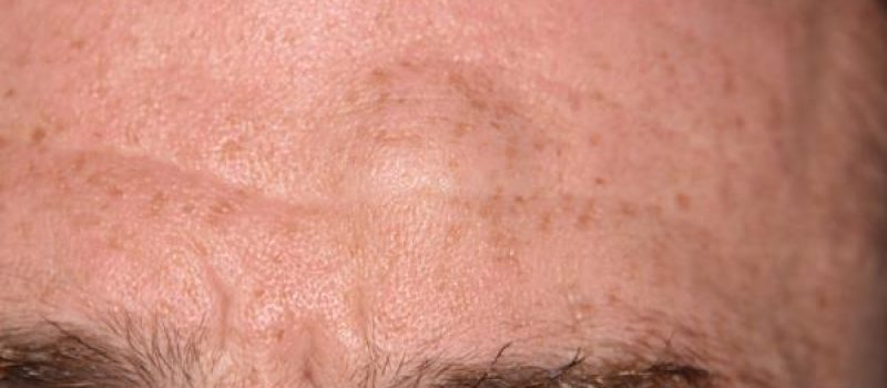
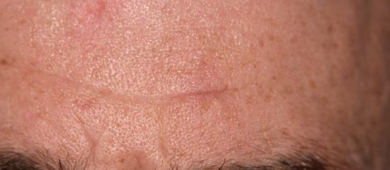
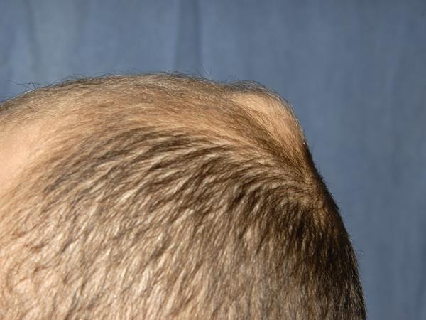
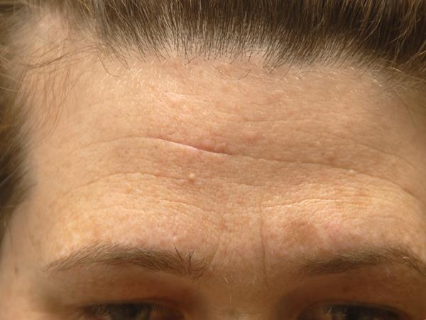
Lipoma 1

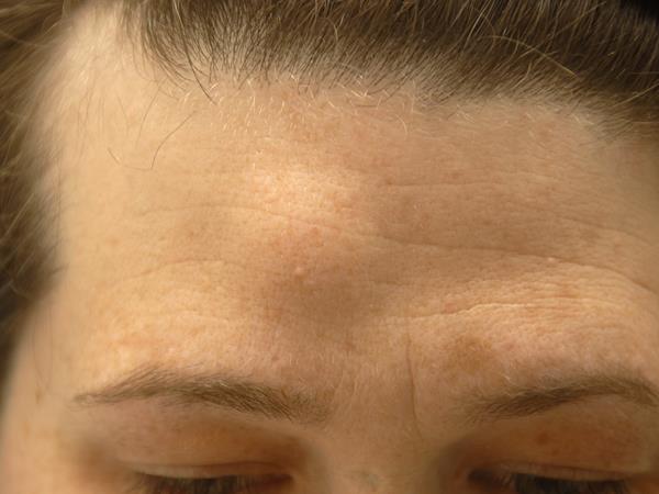
Lipoma forehead in a man.

hurren-lipoma-forehead-1hurren-lipoma-forehead-2

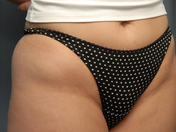
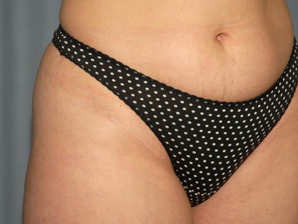
Lipoma 2

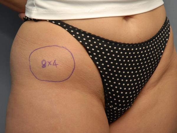
Lipoma right hip.

Lipoma Marked

Scar Close Up After Removal

Lipoma 3

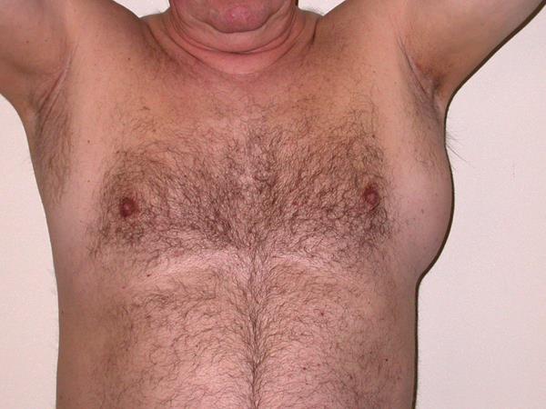
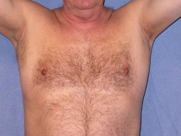
Large lipoma left armpit.

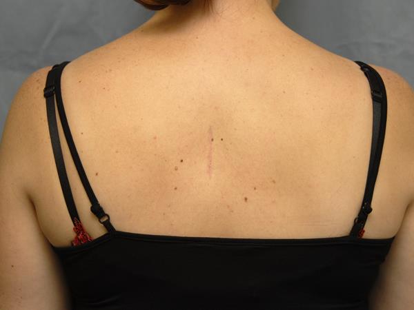
Scar after removal

The Lipoma

Lipoma 4

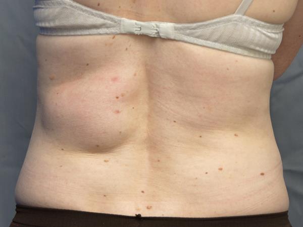
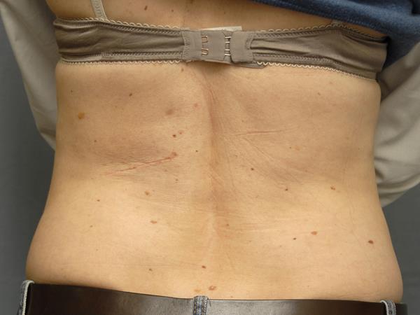
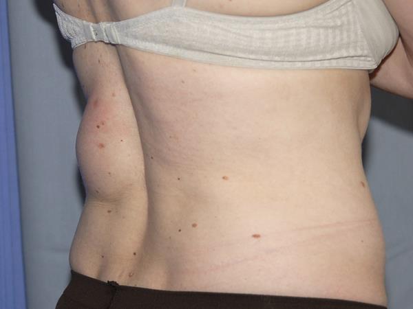
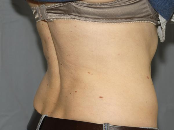
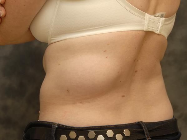
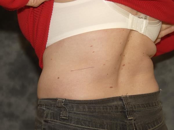
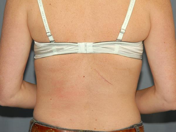
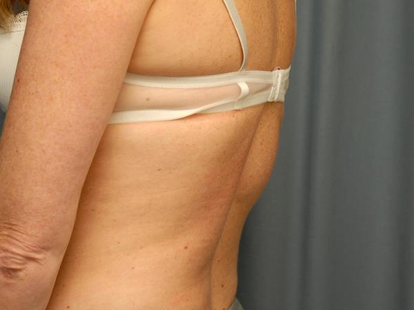
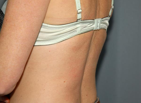
Lipoma left lower back.

Lipoma 5

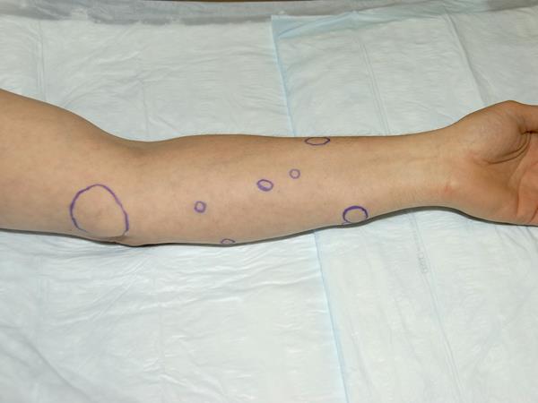
Multiple lipomas on the left arm.

Lipoma 6

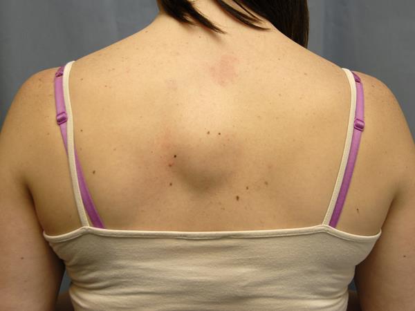
Lipoma middle of back.

Lipoma 7

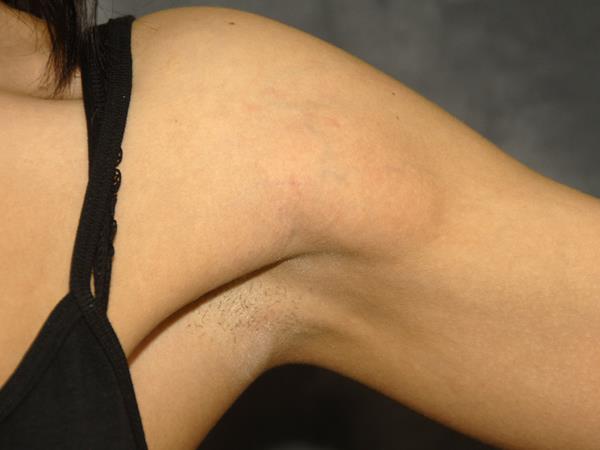
Lipoma front of left arm near armpit.
Lipoma front of left arm near armpit – view of early scar

The Lipoma

Lipoma 8

Lipoma front of left arm near armpit.

The Lipoma

Lipoma 9

Lipoma forehead.

The Lipoma

Lipoma 10

Lipoma forehead.

Lipoma 11

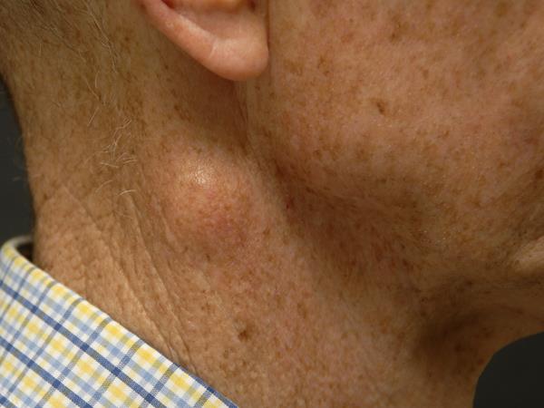
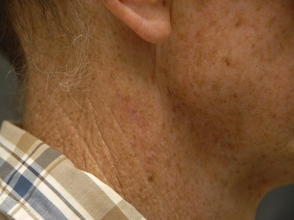
Lipoma right side of neck.

Lipoma 12

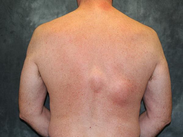
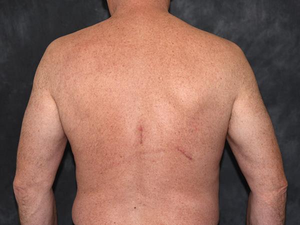
Two lipomas on right side of back.

Lipoma 13

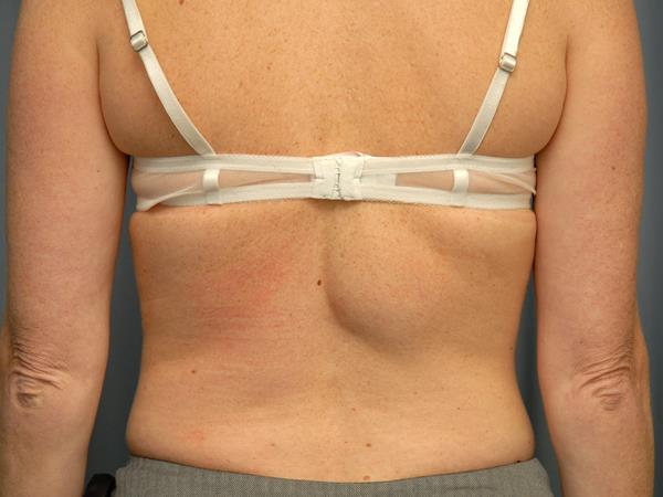
Lipoma right side of back.

Lipoma 14

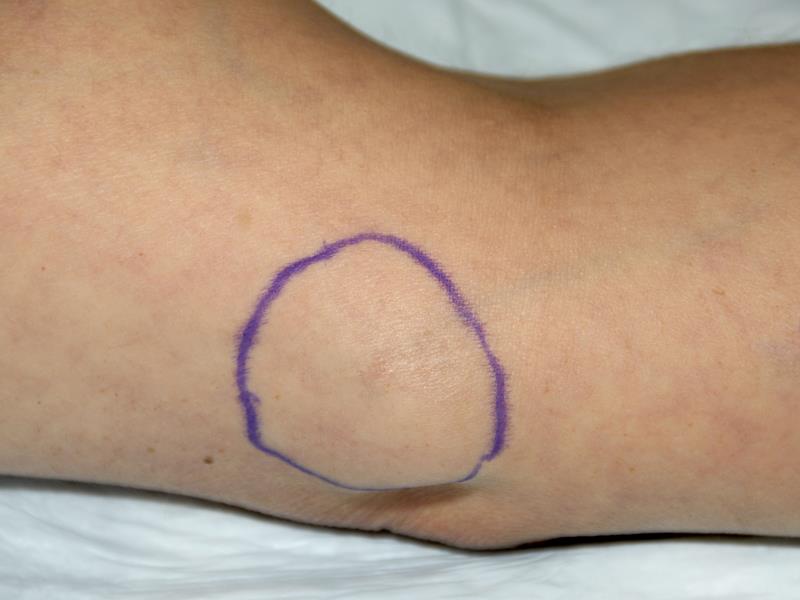
Single lipoma removal from arm